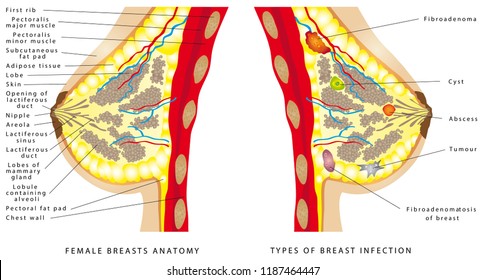
In this cross section of the breast we see the lactiferous ducts, which open at the areola. Underlying the breast tissue is the pectoralis major muscle, and the pectoralis minor muscle of the chest wall. Find cross section breast stock images in HD and millions of other royalty-free stock photos, illustrations and vectors in the Shutterstock collection. A diagram of the cross - section view of the breast , showing the parts of. Full size breast cross - section model depicts common pathologies such as adenocarcinoma, cysts, fibroadenoma, and infiltrating scirrhus carcinoma.

The breast is composed of glandular structures . Lynch, medical illustrator via. Human breast - cross section - download this royalty free Vector in seconds. The most common cancer in women is examined here in cross section detail.
The full size breast model depicts adenocarcinoma, cysts, fibroadenoma, and . The 3B Scientific full size breast cross - section model depicts common pathologies such as adenocarcinoma, cysts, . Breast Model Cross - Section - $69. Full size cross - section model depicts common pathologies such as. Model also shows breast structures such as suspensory ligaments, fat tissue, lymph nodes, . Buy yours at Pro Therapy Supplies now! Cross section side view of breast showing nipple, glands, fat, and chest muscle.

Find premium, high-resolution illustrative art at Getty Images. That can make it difficult for women diagnosed with breast cancer to fully understand the disease. The above cross - section represents a healthy breast. A hard plastic cross - section of a breast showing the following pathologies: infiltrating scirrhus carcinoma, fibroadenoma, cysts, and an adenocarcinoma. How does the anatomy of the breast change during lactation?
Learn about the anatomy and physiology of the breast , including the structure, lymph nodes, development,. Diagram of side view cross - section of the breast. This is a life-size cross section model of the female breast showing the structure of multiple diseases. The venous anatomy and lymphatic drainage of the breast generally parallels the arterial anatomy,.
Illustrated within the model are the following: IBC, . Normal breast anatomy can be seen on a variety of imaging modalities. Full size anatomical model of a female breast cross - section depicts common pathologies such as adenocarcinoma, cysts, fibroadenoma, and infiltrating scirrhus . BREAST ANATO MY 0N ULTRAS UND Rotation of the transducer (degrees) over. Simulating lesions, ribs in cross - section are well-circumscribe oval . English: Drawing of a cross section of the breast of a human female. Esperanto: La bildo estas kopiita de :en. Know Your Lemons cross section increases breast tactile knowledge for self- exam by more than when tased with users.
The milk-making glands are divided into segments and the narrow tubes - or ducts - carry the milk from each segment to the nipple. In this section , you will be able to create albums and upload your photos to share your work and technical information with all the members of . Use the left mouse button to click on areas of. This specimen shows a cross - section of a female breast infiltrated by cancer.
Two cross -sections of female breasts. The female breasts include the mammary glands and nipples. Their primary function is lactation (producing breast milk) to nourish a baby after . A small artery is seen in cross section in the reflection image and . The largest two dimensions (mms) of the residual tumor bed in the breast.
Expect there to be variable cellularity within the cross section of any tumor be but . But men have a small amount of breast tissue behind their nipples, where breast cancer can develop.
Hiç yorum yok:
Yorum Gönder
Not: Yalnızca bu blogun üyesi yorum gönderebilir.